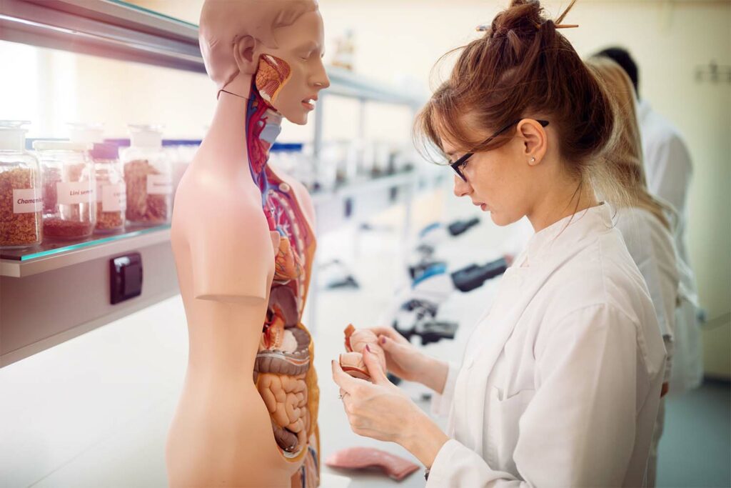
When examining the human body, it can certainly be overwhelming to delve into the microscopic world of cells and tissues, the backbone of anatomy and physiology. More specifically, tissues are groups of like-minded cells that work together toward a common biological task.
The microscopic study of tissues is referred to as histology and is useful to determine how tissues are organized at a cellular level to comprise structures and bodily organs.
What Are the Different Types of Tissues?
Due to the complexity of the human body, along with the wide variety of essential biological functions, many different types of tissues exist. The four main tissue types in the human body are epithelial, muscle, connective, and nervous.
Each respective kind of tissue differs from each other in structure, which subsequently determines its function.
Epithelial
Epithelial tissue is probably the most obvious tissue type — because it covers most of our body in the form of skin! In fact, skin accounts for 16% of the total human body weight and covers approximately 22 square feet on average.
Besides the skin, epithelial tissue comprises our stomach lining, intestines, trachea, and many other passageways in our body that are commonly utilized as a protective layer or for absorbing nutrients.
Within the epithelial tissue distinction exists a handful of other categories of cells characterized by cell shape, size, and layering:
- Squamous epithelial cells are simple, single-layered cells found in areas where diffusion or filtration occurs. Common locations of squamous cells are in the alveoli of the lungs and in capillary walls in blood vessels.
- Cuboidal epithelial cells are like squamous cells in that they are single-layered, but they are a little thicker and possess additional functions that allow them to facilitate secretion or absorption. These cells can be found in kidney tubules and secretory ducts in many different types of glands.
- Columnar epithelial cells are once again single-layered but elongated to provide a larger barrier than either squamous or cuboidal cells. These cells provide protection, such as in the stomach lining against the harsh acidic gastric environment.
- Pseudostratified columnar epithelial cells are columnar cells that possess cilia, a protein structure on the luminal surface of the cell that allows the cell to propel foreign particles or bacteria. For example, the trachea in the respiratory tract uses cilia to direct mucus and inhales foreign entities through the airway.
- Stratified epithelial cells encompass any other kind of epithelial cell that forms in many layers, effectively making a barrier of layered epithelial cells. Some examples include skin, throat, and vaginal linings.
Muscle
Muscle tissue, understandably, is the workhorse of the body. From a simplified viewpoint, muscle tissue allows for movement by receiving neural signals and subsequently contracting. This movement permits us to walk, eat, talk, and even digest food and breathe.
Like epithelial tissue, muscle tissue can be broken down into various other categories, including skeletal, smooth, and cardiac muscle tissue:
- Skeletal muscle tissue makes up what most people think of when they think of muscle tissue, including our biceps and quadriceps muscles, to name a few. A unique characteristic of skeletal muscle is its ability to be under voluntary control, meaning we can use skeletal muscle whenever we want.
- Cardiac tissue has a specialized purpose and functions to pump blood throughout the body via the heart. Unlike skeletal muscle, cardiac muscle operates mostly involuntarily, although we can certainly do and think about things to increase or decrease its degree of activity.
- Smooth muscle cell tissue is a less obvious large source of muscle tissue. Smooth muscle cells line blood vessels and our gastrointestinal tract and serve to promote the movement of fluids, food, blood, or whatever else needs to be transported throughout our body. Once again, it functions involuntarily but can contract and relax as a result of either neural parasympathetic stimulation or chemical stimulation.
Connective
Connective tissue is an often-overlooked tissue type. You might be surprised to learn that connective tissue is the most abundant type of tissue in the human body. As its name suggests, it works to connect other cells and tissues together, and it can be found nearly everywhere in our body. Typically, we can find connective tissue in bones, cartilage, adipose, and collagen.
The importance of connective tissue is not always readily discernible. However, essential bodily functions such as storing fat, protecting muscle and bone, and providing structure to other tissues and organs in the body would not be possible without it.
Similar to previous tissue types, connective tissue can be split into different categories: loose, dense, and specialized. Loose connective tissue can be thought of as the “glue” that holds everything in place, while dense makes up tendons and ligaments with high-density collagen. Specialized connective tissue can be found in adipose, cartilage, bone, blood, and even lymph fluid.
Other cells are included in the connective tissue category, such as white blood cells, plasma, mast cells, and macrophages. Moreover, blood is considered a special, atypical connective tissue. Blood is composed of suspended cells in a fluid matrix known as plasma and contains red blood cells, plasma proteins, and hormones, to name a few.
Nervous
Neural tissue maintains our body’s communication system. Electrical signals are transduced via neurons to perform functions that help us walk, think, and allow our organs to operate without conscious thought. Neurons are cells themselves and contain specialized features that allow them to receive and facilitate this type of communication throughout the body.
There are two types of neural cells in our neural tissue: neurons and glia. Neurons are the main communicative cells that are responsible for electrical signal transduction, while glial cells provide support by regulating the health of neural cells and providing protection for the neurons’ axons.
How Do We Prepare a Tissue Sample?
The histopathological examination begins by collecting a tissue sample. This can be through surgery, biopsy, or autopsy. Following collection, a tissue sample will have to be dissected and fixated, a process that prevents tissue decay and ultimately preserves it for long-term inspection.
Depending on the type of tissue, this can be achieved via chemical fixation utilizing a compound called formalin or through freezing the tissue. Formalin effectively preserves tissue by inactivating enzymes and preventing cell autolysis and self-digestion.
Processing the tissue sample can be time and skill intensive and is commonly performed by a histotechnician or histotechnologist, both specially trained laboratory technicians. Through preparation with alcohol to remove water followed by wax infiltration, tissue samples can be sliced into cross sections that will allow them to be observed under a microscope.
Stain can also be applied with one or more pigments, again depending on the tissue type. The purpose of staining is to highlight important cellular components against a highly visible contrast.
A commonly used stain, hematoxylin, and eosin (H&E), acts as a combination of basic and acidic dye, which allows better visualization of cell structures. Hematoxylin is basophilic and reacts strongly with anionic cell structures such as DNA and RNA. Alternatively, eosin is acidophilic and reacts strongly with cationic structures such as collagen and many proteins.
What Are Some Applications for Histology?
Histology is essential to and applied in numerous scientific and medical fields. Understanding the anatomical and surgical pathology of tissue can provide invaluable information regarding disease progression.
Medical Histology
In clinical medicine, medical histology, sometimes called histopathology, refers to the microscopic examination of a tissue specimen by a pathologist, a professional that studies the causes of disease. A pathologist is trained to formulate a pathology report, an often-essential component contributing to a medical diagnosis.
Cancer is often diagnosed with histopathological examination. Commonly, this process directs what treatment protocol is prescribed based on the characteristics of the cancer cell. Sometimes immunohistology, a process that involves visualizing a protein or biomarker in tissue using antibodies, is used in combination with a cancer diagnosis to further characterize what treatment will be most effective in patients.
Histopathology is even implicated towards the end of treating a cancer diagnosis by indicating surgical margins of cancer cells, suggesting how well the cancer has been treated.
Summary
In conclusion, we can see that histology is a vast but essential field of science and medicine. From its inception in the early 1800s, histology continues to guide and advance our understanding of human and animal tissue.
Coming up with quality biospecimens to advance research can sometimes be challenging, but at research corporations such as iProcess, obtaining a quote is easy and ensures the highest quality samples are delivered to you to meet your research deadlines.
iProcess supplies specimens including tissues, blood, plasma, serum, urine, stool, and swab samples to many companies ranging from start-ups to major pharmaceutical, life science, diagnostic, and research organizations.
Sources:

